|
EYE
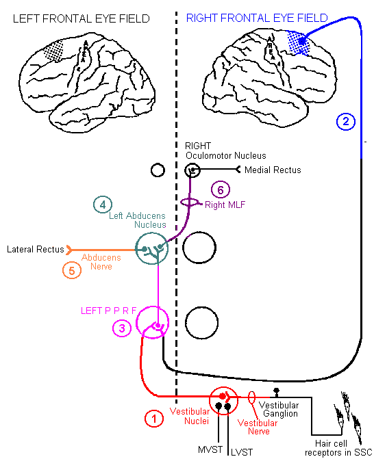
MOVEMENTS AT A GLANCE (diagram below)
|
|

staggering to the RIGHT;
LEFT nystagmus
|

(575k) (287k)
|
|

both eyes deviated to the RIGHT
(away from the hemiplegia); inability to
voluntarily turn eyes past the midline to the LEFT;
NO muscle atrophy; NO diploplia
|
(574k) (287k)
(660k) (330k)
|
|

both eyes deviated to the RIGHT
(toward the hemiplegia); inability to voluntarily
turn eyes past the midline to the LEFT; NO muscle
atrophy; NO diploplia
|
(624k) (312k)
(355k) (178k)
|
|

both eyes deviated to the RIGHT;
inability to voluntarily turn eyes past the midline
to the LEFT; ATROPHY of the LEFT lateral rectus but
not the right medial rectus; NO diploplia
|
(337k) (170k)
(559k) (279k)
|
|

LEFT eye deviated medially
Diploplia (corrected by turning head to LEFT)
ATROPHY of the LEFT lateral rectus
|
(398k) (199k)
(508k) (254k)
|
|

RIGHT eye deviated laterally
Diploplia (corrected by turning head to the
LEFT)
NO atrophy of the right medial rectus (can still
contract the right medial rectus during
convergence; convergence center is rostral to the
occulomotor complex)
|
(637k) (319k)
(280k) (140k)
(473k) (237k)
|