Point
1
Intro
 So, here comes the
information about two point discrimination, vibration and
conscious proprioception (very important stuff!!) over
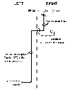
the central process of the alpha-beta axon. As the central
process of the alpha-beta axon approaches the spinal cord it
travels in what is called the medial division of the
dorsal root (this medial group of fibers will be
contrasted with other central processes that lie laterally
in the dorsal root). Once in the dorsal funiculus, the
alpha-beta axon takes off for the medulla, where it
synapses. The medulla is the most caudal part of the
brainstem (midbrain, pons, medulla), and it lies immediately
rostral to the spinal cord. Remember--there has been no
synapse in the dorsal root OR spinal cord. Also, the
axon does NOT CROSS IN THE SPINAL CORD!! It
terminates in the medulla on the same side
(IPSILATERAL; ipsi = L., same; latus = side) as its
cell body. Cells in nucleus gracilis and cuneatus
project to the thalamus, the information is then relayed to
somatosensory cortex for perception. More on this later!!
Let’s stick to the spinal cord for now.
So, here comes the
information about two point discrimination, vibration and
conscious proprioception (very important stuff!!) over
the central process of the alpha-beta axon. As the central
process of the alpha-beta axon approaches the spinal cord it
travels in what is called the medial division of the
dorsal root (this medial group of fibers will be
contrasted with other central processes that lie laterally
in the dorsal root). Once in the dorsal funiculus, the
alpha-beta axon takes off for the medulla, where it
synapses. The medulla is the most caudal part of the
brainstem (midbrain, pons, medulla), and it lies immediately
rostral to the spinal cord. Remember--there has been no
synapse in the dorsal root OR spinal cord. Also, the
axon does NOT CROSS IN THE SPINAL CORD!! It
terminates in the medulla on the same side
(IPSILATERAL; ipsi = L., same; latus = side) as its
cell body. Cells in nucleus gracilis and cuneatus
project to the thalamus, the information is then relayed to
somatosensory cortex for perception. More on this later!!
Let’s stick to the spinal cord for now.
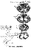
 There are two components to the dorsal column
system, called fasciculus gracilis and fasciculus
cuneatus (fasciculus = L., little bundle; gracilis =
slender; cuneatus = wedge). The central process of the
alpha-beta fiber travels within the fasciculus gracilis if
it arises from dorsal root ganglia T7 and below. In
contrast, if the central process of the alpha-beta
fiber arises from cells in dorsal roots T6 and above
(toward your head), it is part of fasciculus cuneatus
(THINK: GRACILIS = LEG AND CUNEATUS = ARM).
There are two components to the dorsal column
system, called fasciculus gracilis and fasciculus
cuneatus (fasciculus = L., little bundle; gracilis =
slender; cuneatus = wedge). The central process of the
alpha-beta fiber travels within the fasciculus gracilis if
it arises from dorsal root ganglia T7 and below. In
contrast, if the central process of the alpha-beta
fiber arises from cells in dorsal roots T6 and above
(toward your head), it is part of fasciculus cuneatus
(THINK: GRACILIS = LEG AND CUNEATUS = ARM).
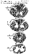 Fasciculus gracilis and
fasciculus cuneatus are thus comprised of the alpha-beta
axons whose cell bodies lie in IPSILATERAL DORSAL ROOT
GANGLIA. That is, the cell bodies are on the SAME
SIDE as the fasciculi. I have mentioned that fibers in
the dorsal column system DO NOT CROSS in the spinal
cord and eventually synapse in the medulla. While we will
cover the medulla later in the course, you might like to
know that axons in fasciculus gracilis terminate in the
ipsilateral (to the fasciculus) nucleus gracilis,
while fibers in fasciculus cuneatus synapse in ipsilateral
nucleus cuneatus (big surprise).
Fasciculus gracilis and
fasciculus cuneatus are thus comprised of the alpha-beta
axons whose cell bodies lie in IPSILATERAL DORSAL ROOT
GANGLIA. That is, the cell bodies are on the SAME
SIDE as the fasciculi. I have mentioned that fibers in
the dorsal column system DO NOT CROSS in the spinal
cord and eventually synapse in the medulla. While we will
cover the medulla later in the course, you might like to
know that axons in fasciculus gracilis terminate in the
ipsilateral (to the fasciculus) nucleus gracilis,
while fibers in fasciculus cuneatus synapse in ipsilateral
nucleus cuneatus (big surprise).
The fasciculus gracilis contains fibers from spinal cord levels lower than fasciculus cuneatus, and fasciculus gracilis lies MEDIAL to fasciculus cuneatus. This lower = medial spatial relationship holds not only for the two fasciculi, but also for the individual fibers in each fasciculi. For example, the most medially placed fiber in fasciculus gracilis arises from the coccygeal dorsal root and the most laterally placed arises from the T7 dorsal root. In the fasciculus cuneatus, the most medially placed fiber arises from dorsal root T6 and the most lateral arises from dorsal root C2 (remember from Gross Anatomy that C1 is purely motor, and therefore does not have a dorsal root ganglion?!!).