Point
6
Intro
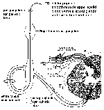 The preganglionic
sympathetics pass to the sympathetic trunk via the white
communicating rami. They can synapse in the autonomic trunk
(paravertebral ganglia), go up or down and synapse, or go
through to comprise the splanchnics. Preganglionic
sympathetics from spinal levels T1 to T5 ascend to the
superior cervical ganglion, but most of these fibers arise
from T1. Cells in the superior
cervical ganglion (which receive their main drive from cells
in the lateral horn at spinal level T1) then innervate, via
postganglionic sympathetics, the smooth muscle of the
dilator pupillae, the smooth muscle of the upper eye lid,
the blood vessels, sweat glands, and hair of the head and
face. A LESION at spinal level T1, either in the
spinal cord or the ventral root, interrupts the sympathetic
drive to these structures. This results in what is called
HORNER'S SYNDROME. On the side
IPSILATERAL to the spinal T1 lesion there is a
drooping eyelid (PTOSIS), a constricted pupil
(MIOSIS; remember, the boring parasympathetics are
"in charge"), and a flushed (vasodilatation, since
sympathetics to the skin vasoconstrict) and dry face.
The preganglionic
sympathetics pass to the sympathetic trunk via the white
communicating rami. They can synapse in the autonomic trunk
(paravertebral ganglia), go up or down and synapse, or go
through to comprise the splanchnics. Preganglionic
sympathetics from spinal levels T1 to T5 ascend to the
superior cervical ganglion, but most of these fibers arise
from T1. Cells in the superior
cervical ganglion (which receive their main drive from cells
in the lateral horn at spinal level T1) then innervate, via
postganglionic sympathetics, the smooth muscle of the
dilator pupillae, the smooth muscle of the upper eye lid,
the blood vessels, sweat glands, and hair of the head and
face. A LESION at spinal level T1, either in the
spinal cord or the ventral root, interrupts the sympathetic
drive to these structures. This results in what is called
HORNER'S SYNDROME. On the side
IPSILATERAL to the spinal T1 lesion there is a
drooping eyelid (PTOSIS), a constricted pupil
(MIOSIS; remember, the boring parasympathetics are
"in charge"), and a flushed (vasodilatation, since
sympathetics to the skin vasoconstrict) and dry face.